Exosomes are membrane-bound vesicles released by cells that mediate intercellular communication and have potential for clinical applications such as monitoring disease progression and drug delivery. However, their broad size distribution, including larger vesicles, challenges traditional detection methods, which often fail to capture the true size distribution. Nanocoulter™, with its precise and reproducible single-particle detection, offers new possibilities for advancing extracellular vesicle (EV) research.
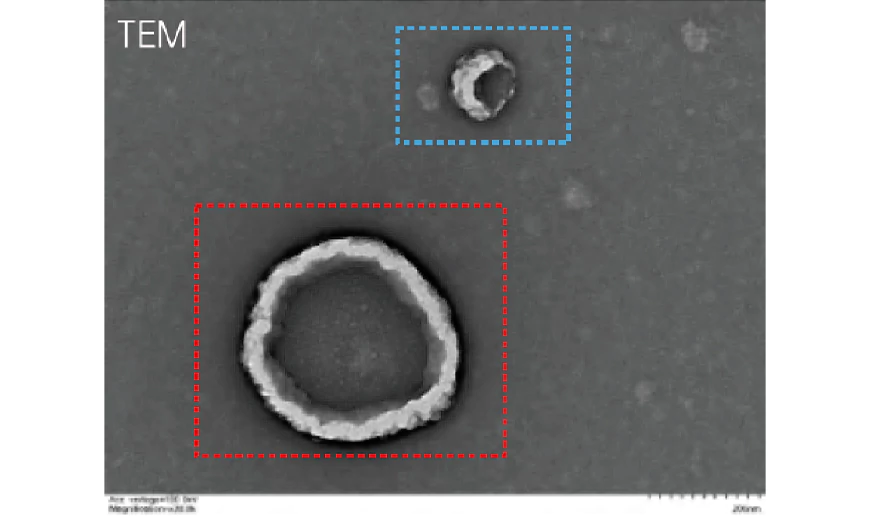
Comparable to Electron Microscopy in Particle Size Detection
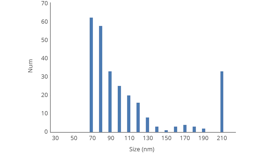
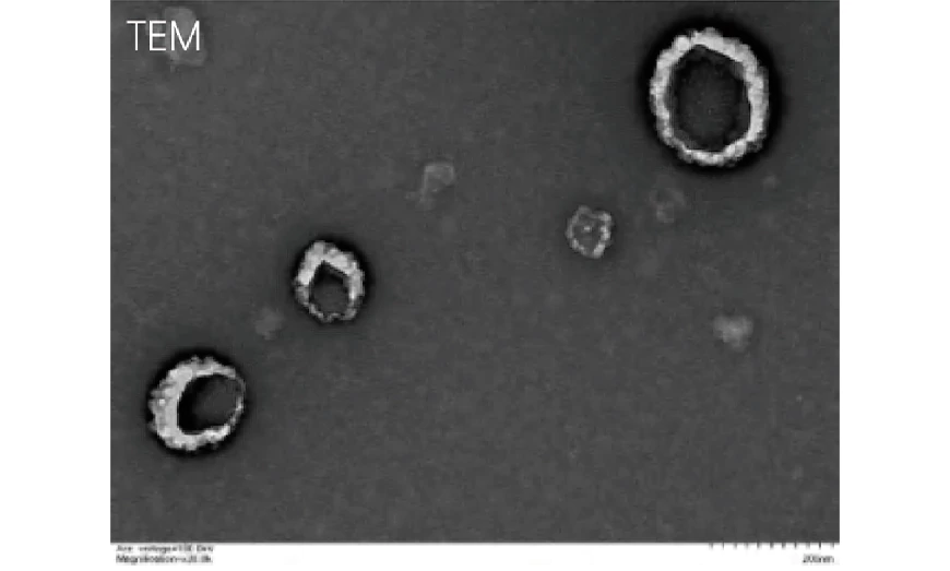
The Nanocoulter™ Single-Particle Analyzer delivers exceptionally high particle size resolution—comparable to electron microscopy—while enabling quantitative analysis across a broad size distribution. It accurately measures both small and large particles in heterogeneous samples, providing detailed insights into the proportion of particles within each size range.
The graph below shows the analysis of an exosome sample measured using Nanocoulter™. A clear bimodal distribution is visible, with the majority of particles falling between 70–90 nm, closely matching exosome size data obtained from electron microscopy studies.
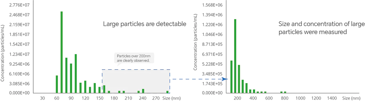
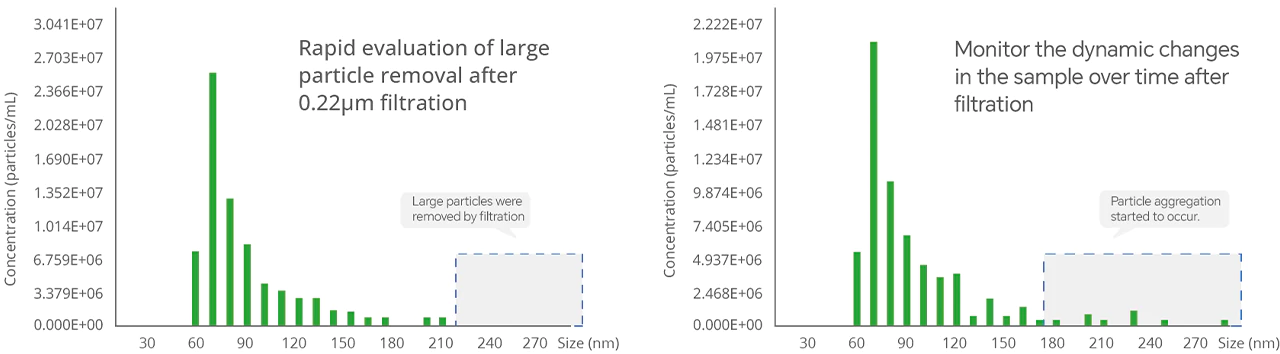
EVs Size Distribution and Aggregation Detection
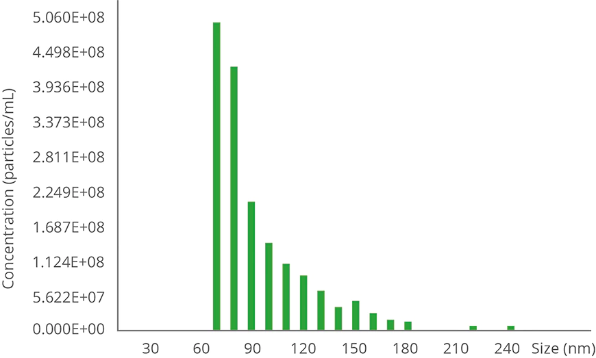
NanoCoulter™ enables precise measurement of large vesicles in EVs samples. By utilizing nanochips with different measurement ranges, it can determine particle concentrations across various size ranges. With its ultra-high sensitivity for single-particle detection, NanoCoulter™ swiftly identifies subtle changes before and after sample treatment, significantly accelerating the research and development of EVs.
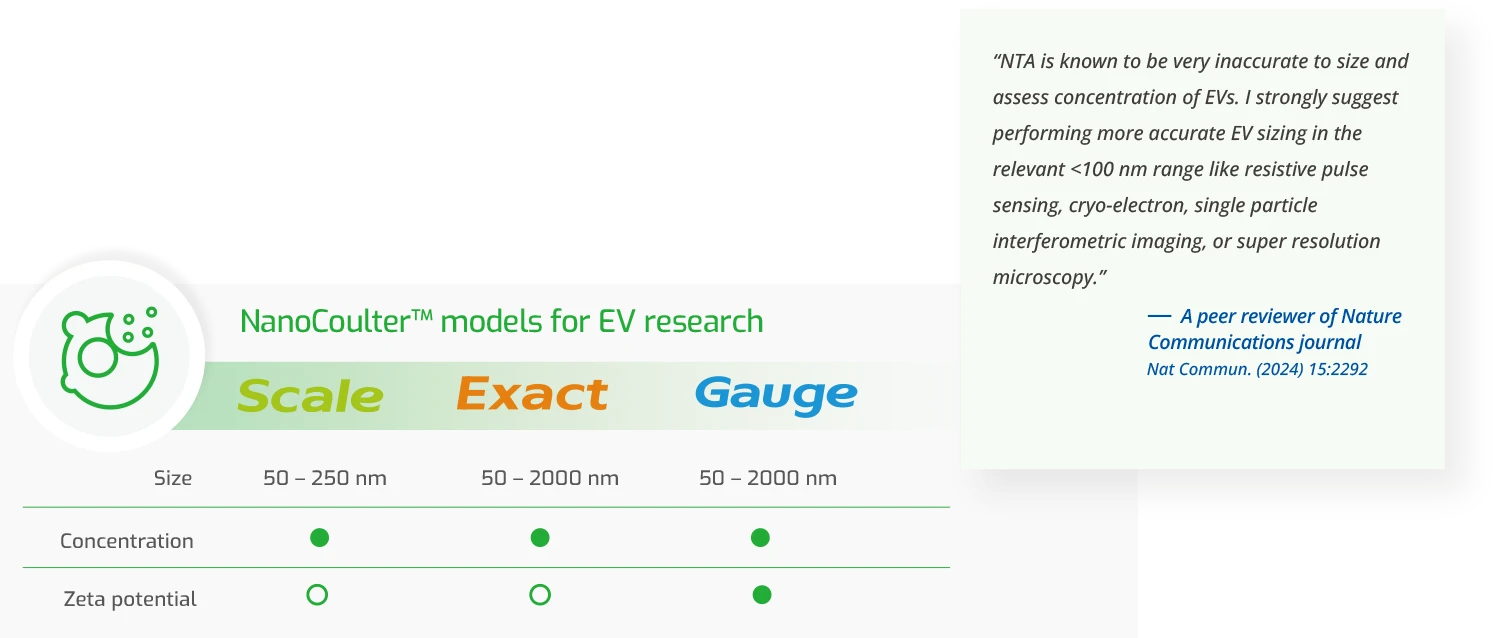
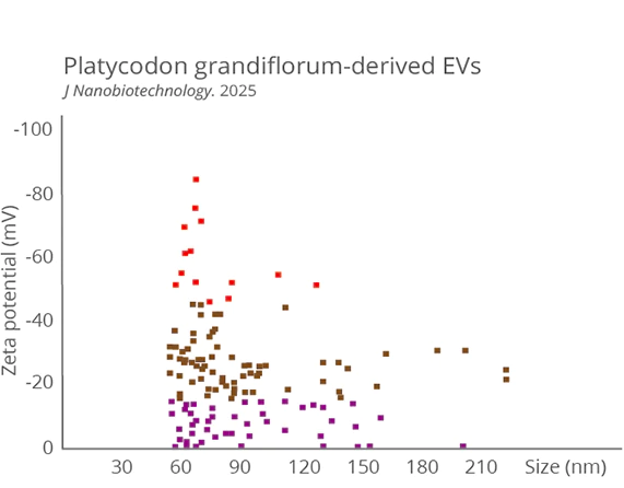
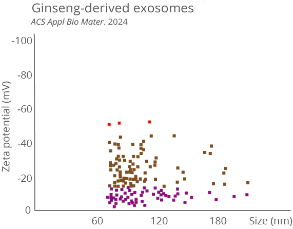
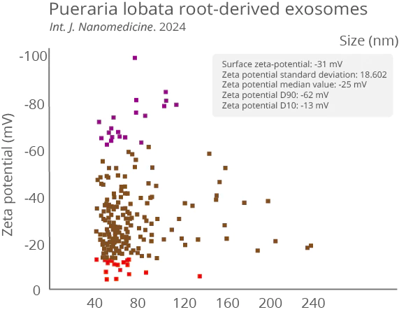
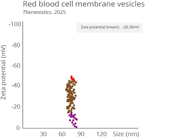
EV Zeta Potential Characterization
Zeta potential is a key parameter for evaluating EV stability, surface charge characteristics, and in vivo interactions. It directly influences adsorption, targeted delivery, and circulation time within biological systems. Traditional optical methods (DLS/NTA) can only provide an ensemble-averaged potential, failing to capture sample heterogeneity. NanoCoulter™ enables single-particle zeta potential measurement, offering precise insights into EV population heterogeneity—advancing research and applications in EV-based therapeutics.
Investigation of EV Isolation Methods
EVs purification often requires multiple steps. NanoCoulter™ quickly and accurately evaluates the impact of different methods on EV size and concentration.
Characterization of Engineered Exosomes
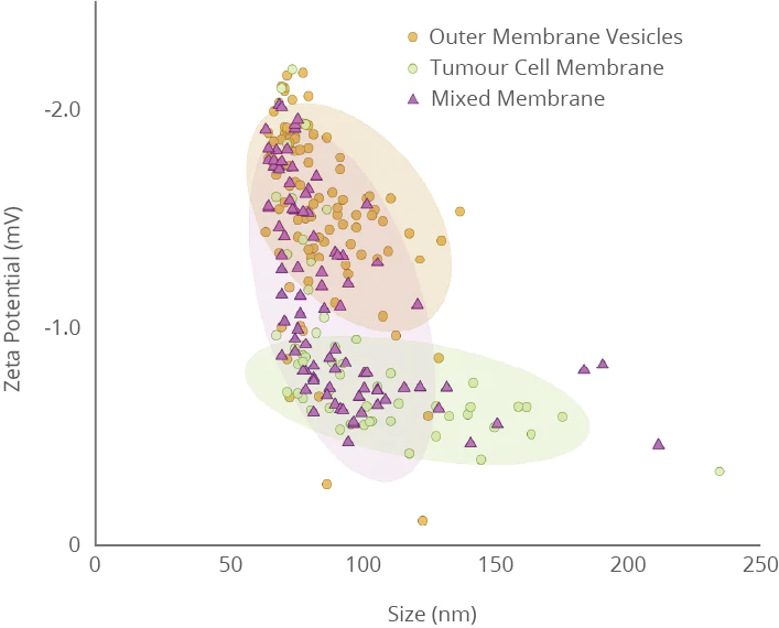
Characterization of fused exosomes
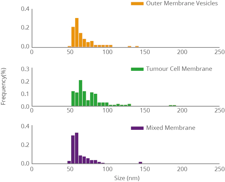
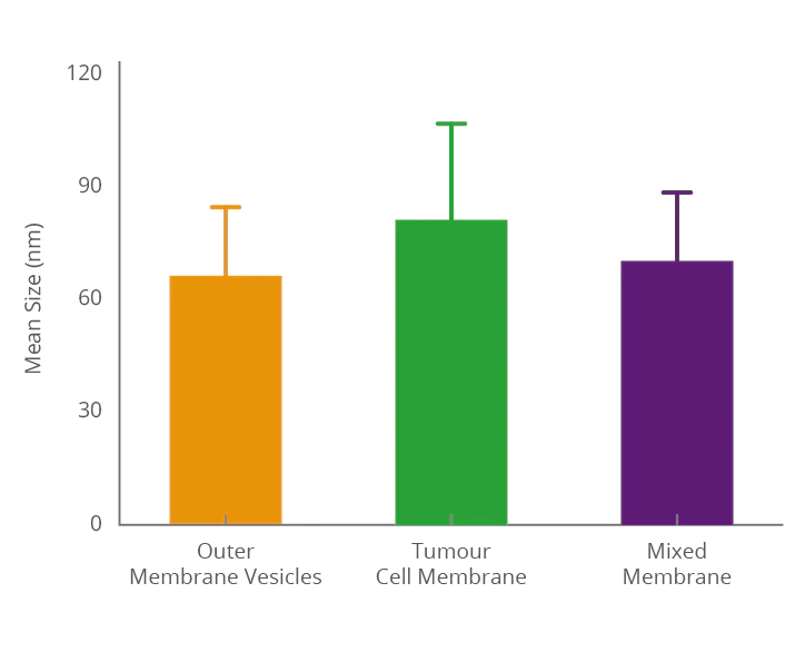
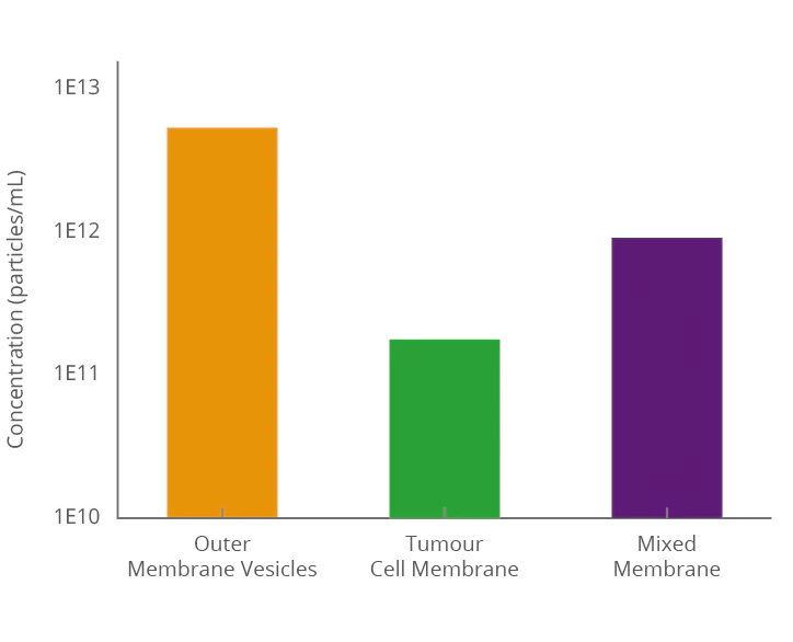
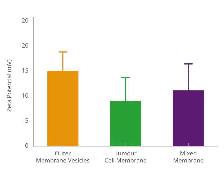
Fusing bacterial outer membrane vesicles with cancer cell exosome membranes enables the hybrid vesicles to simultaneously inherit biological functions from both bacterial and cancer cell membranes, creating a multifunctional delivery system. Precise characterization of the fused exosomes using NanoCoulter™ allows clear visualization of key parameters such as particle size distribution, concentration, and zeta potential, thereby confirming membrane fusion efficiency and providing reliable data support for the development of targeted drug or vaccine carriers.
Characterization of Drug-loaded Exosomes
During the development and quality assessment of drug-loaded exosomes, NanoCoulter™ offers a robust platform for single-particle characterization. It enables accurate quantification of key physicochemical parameters, including particle size distribution, concentration, and zeta potential. These measurements facilitate the evaluation of drug loading efficiency, particle homogeneity, and colloidal stability. Unlike conventional optical techniques, NanoCoulter™ does not rely on fluorescence labeling or refractive index assumptions, thereby providing higher analytical fidelity for irregularly shaped particles and complex sample matrices. This advantage makes it particularly valuable in process optimization, batch-to-batch consistency assessment, and the design of targeted delivery systems.
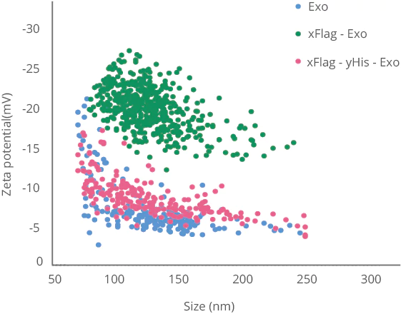
Exosome surfaces were modified with (x)Flag-tag and (y)His-tag, respectively, to modulate the stability and targeting ability of EV-based drug delivery vehicles. NanoCoulter™ enables clear characterization of the changes in zeta potential before and after surface modification.
The potential change of exosomes modified with protein tags
Optimization of Exosome Cell Culture
During the cell culture process for exosome production, isolation and enrichment are typically required before quantification. If the purity is insufficient or the concentration is too low, the material often fails to meet the needs of downstream research and development, necessitating a repeat of the culture process—resulting in significant time loss. Therefore, researchers are in urgent need of an accurate, reliable, and rapid detection method to monitor exosome secretion levels in real time during cell culture, in order to improve experimental efficiency.
The NanoCoulter™ Single-Particle Analyzer is specifically designed for such applications, enabling more efficient and precise exosome research—with twice the results for half the effort.
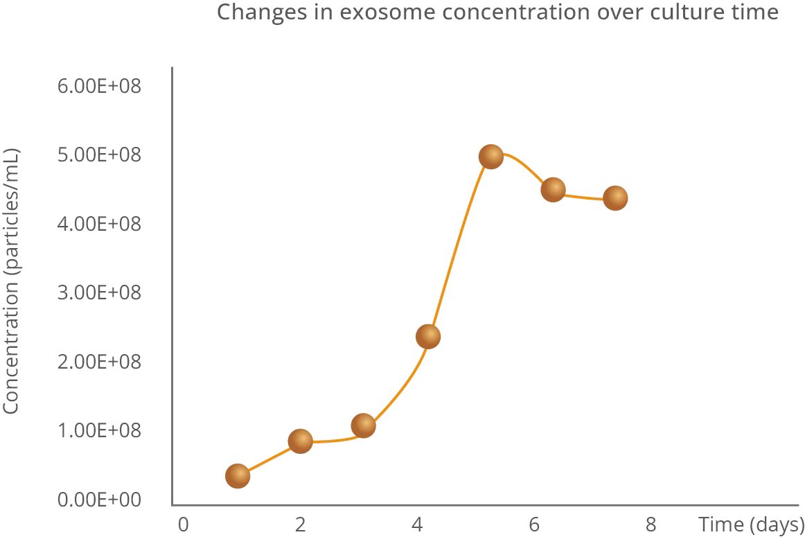
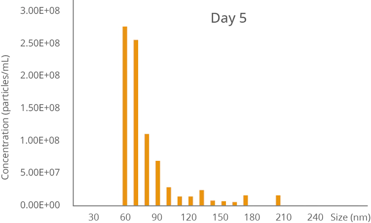
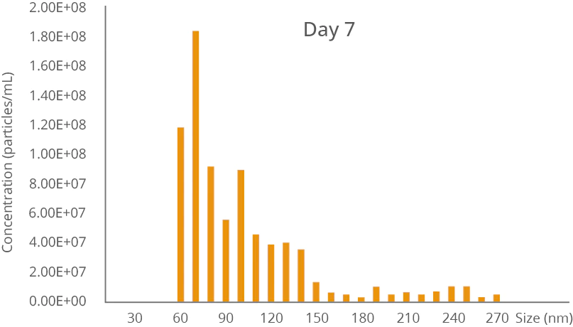
During continuous cell culture over 7 days, exosome secretion was tracked, revealing the peak concentration on day 5.
Purity Assessment of EVs
Rapid quantitative assessment of EV or exosome purity by measuring the particle concentration of treated and untreated samples by detergent-lysis.
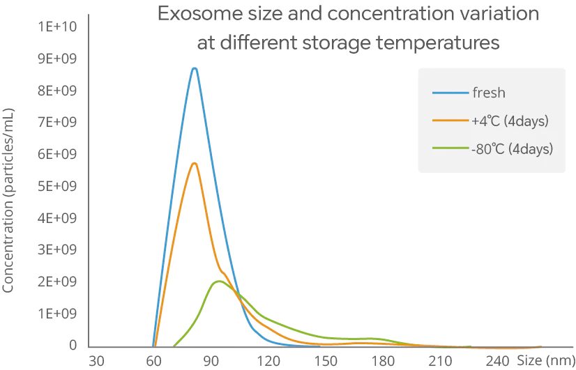
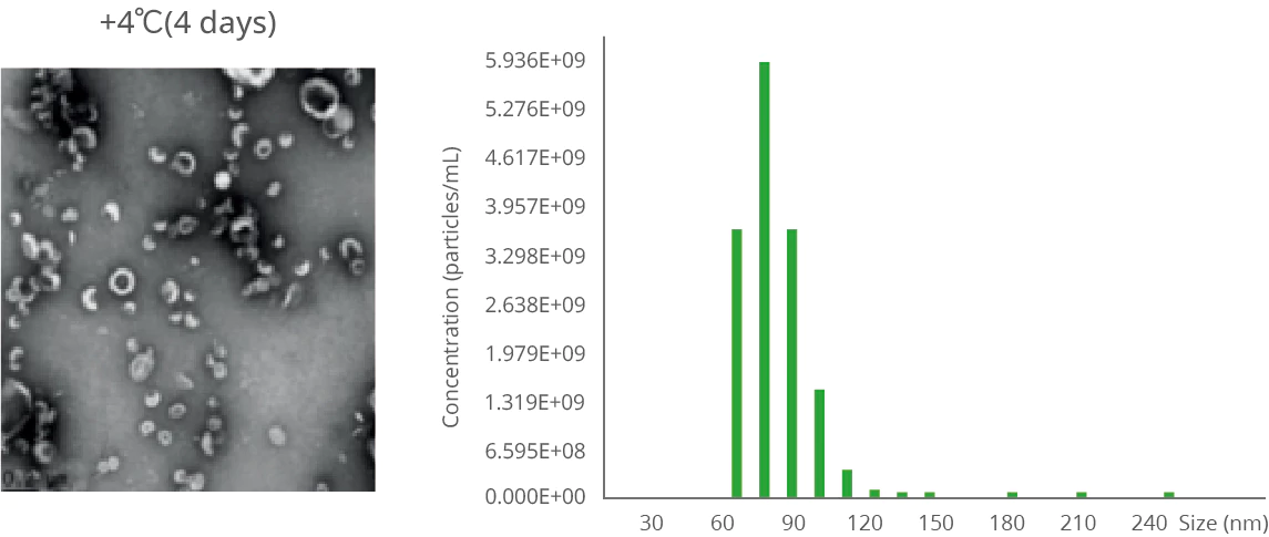
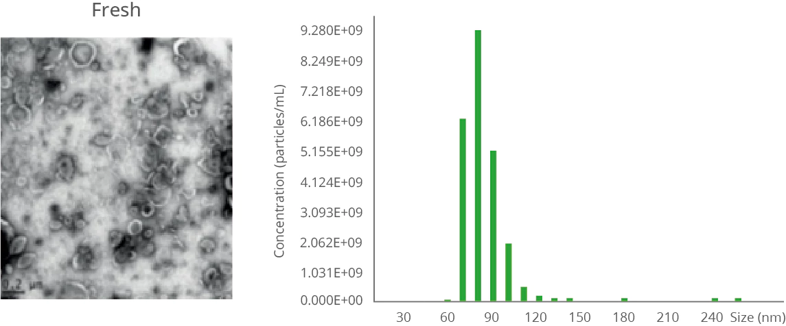
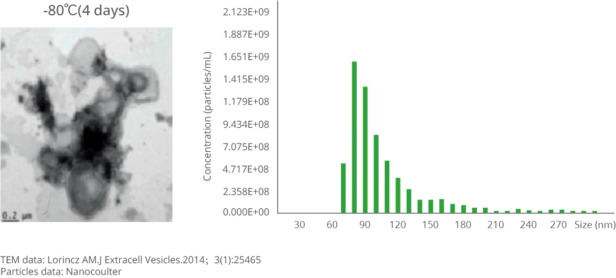
Extracted exosome samples should be stored at 4°C for short-term preservation and at -80°C for long-term storage. However, regardless of the storage conditions, exosome samples undergo varying degrees of change. Researchers conducted a comparative study to analyze changes in newly extracted exosomes after 4 days of storage at 4°C and -80°C. The results showed that after 4 days at 4°C, the particle size of the exosomes changed only slightly, whereas after 4 days at -80°C, the particle size and morphology of the exosomes changed significantly, with a marked increase in larger particles. The experiment was repeated using a NanoCoulter particle analyzer, and the measured data matched previous findings precisely, demonstrating that the NanoCoulter particle analyzer can accurately monitor the impact of storage temperature on exosomes.